

Two-Dimensional Echocardiography (2D Echo Color Doppler) is a safe, painless, and non-invasive ultrasound test that produces detailed images of the heart, heart valves, and major blood vessels. It helps cardiologists evaluate the structure and function of the heart with high accuracy.
Advanced 2D Echo Color Doppler examination for accurate evaluation of heart chambers, valves, pumping function, and blood flow using modern cardiac imaging technology.
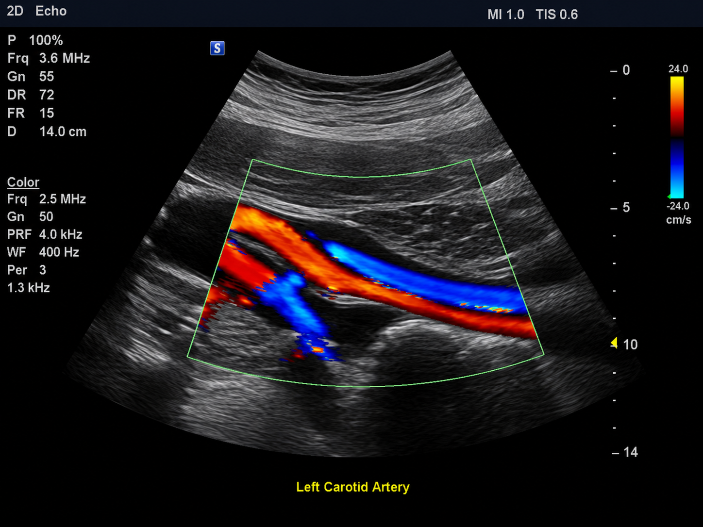
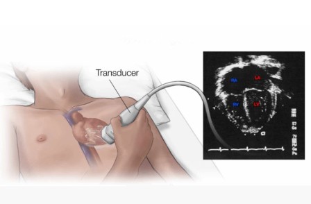
Two-Dimensional Echocardiography (2D Echo) provides detailed real-time images of the heart, surrounding cardiac structures, and major blood vessels. During the examination, a handheld device called a transducer sends high-frequency sound waves toward the heart and receives the returning echoes to create detailed images.
The images are displayed in both black & white and color, allowing cardiologists to assess the size of the heart, movement of the heart walls, valve function, and pumping efficiency. The test is completely painless and does not involve radiation.
A 2D Echo Color Doppler helps cardiologists evaluate the structure, function, and blood flow of the heart for accurate diagnosis and treatment planning.
Measures the size and function of each chamber of the heart.
Evaluates the pumping strength and movement of the heart muscle.
Color Doppler shows the direction and speed of blood flow through the heart.
Detects valve narrowing, leakage, and other structural abnormalities.
Dr. Shripad V. Khairnar provides comprehensive 2D Echocardiography and Color Doppler evaluation to assess the structure and functioning of the heart. The test helps evaluate heart chambers, valves, pumping function and blood flow, supporting accurate diagnosis and appropriate cardiac treatment.