

Trans Esophageal Echocardiography (TEE) is an advanced ultrasound imaging test that produces highly detailed, real-time images of the heart and its internal structures. Since the ultrasound probe is positioned inside the esophagus, TEE provides clearer images than a standard echocardiogram, helping cardiologists diagnose complex heart conditions with greater accuracy.
Advanced Transesophageal Echocardiography (TEE) for accurate diagnosis of heart valve disorders, blood clots, structural heart disease, and cardiac abnormalities.
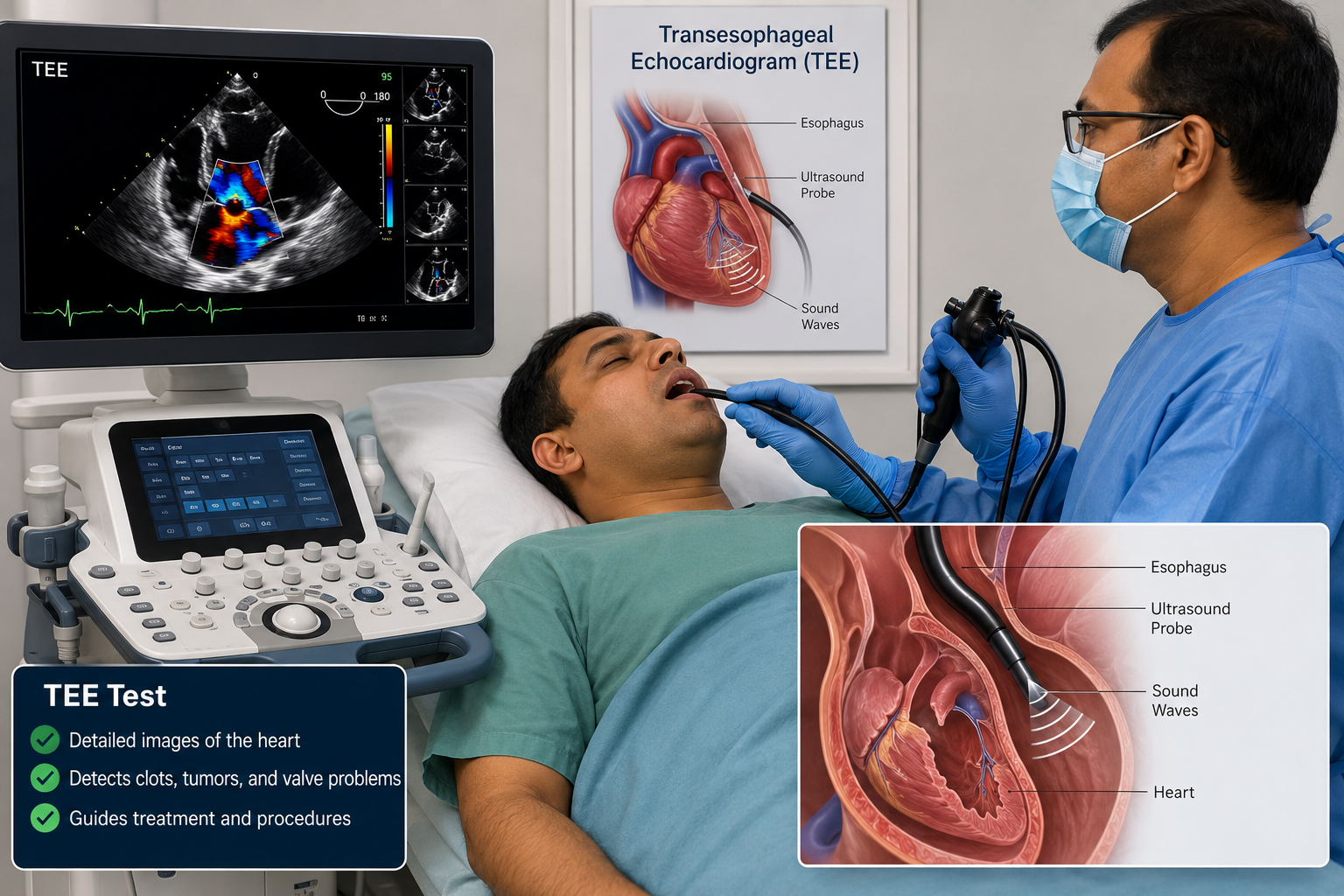
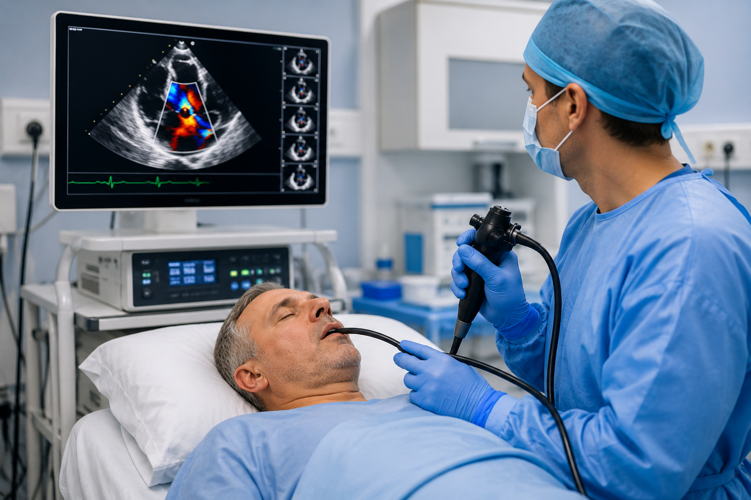
A Transesophageal Echocardiogram (TEE) is an advanced ultrasound imaging test that uses sound waves to produce highly detailed, real-time images of the heart. Because the ultrasound probe is placed inside the esophagus, which lies directly behind the heart, TEE provides clearer and more detailed images than a standard echocardiogram performed through the chest.
TEE allows cardiologists to evaluate the heart chambers, valves, blood vessels, and surrounding structures with exceptional clarity, making it an important diagnostic tool for many complex heart conditions.
TEE provides highly detailed images of the heart and is recommended when more precise evaluation is required than a standard echocardiogram can provide.
Evaluates heart valves for narrowing, leakage, infection, and other abnormalities.
Identifies blood clots inside the heart before procedures such as atrial fibrillation treatment.
Detects abnormalities in heart chambers, walls, and major blood vessels.
Assists cardiologists in accurately guiding catheters during diagnostic and treatment procedures.
Transesophageal Echocardiography (TEE) provides exceptionally clear images of the heart, allowing cardiologists to diagnose complex cardiac conditions with greater precision. It is especially useful when a standard echocardiogram cannot provide enough detail.
Your cardiologist may recommend a Transesophageal Echocardiogram when detailed evaluation of your heart is required beyond a standard echocardiogram.
Evaluates damaged, narrowed, leaking, or infected heart valves with exceptional image quality.
Detects blood clots within the heart before cardioversion or atrial fibrillation treatment.
Provides important information for patients with abnormal heart rhythms and irregular heartbeats.
Identifies abnormalities involving heart chambers, septum, and major blood vessels.
Provides real-time imaging to accurately guide catheter-based cardiac procedures.
Recommended when standard transthoracic echocardiography does not provide sufficient diagnostic information.
TEE offers superior visualization of the heart, helping cardiologists diagnose heart disease accurately and make better treatment decisions.
Produces highly detailed images of the heart compared to standard echocardiography.
Improves detection of heart valve disease, blood clots, and structural abnormalities.
Helps cardiologists plan and safely perform complex heart procedures.
Provides reliable information for personalized treatment and improved patient outcomes.
A Transesophageal Echocardiography (TEE) test is performed by gently passing a thin, flexible ultrasound probe through the mouth into the esophagus. Since the esophagus lies directly behind the heart, the probe captures exceptionally clear, real-time images of the heart without interference from the ribs or lungs. The procedure is carried out under local throat anesthesia and mild sedation to ensure maximum comfort.
Transesophageal Echocardiography provides detailed cardiac imaging, helping cardiologists diagnose heart conditions accurately and plan effective treatment with confidence.
Produces superior-quality images of the heart compared to a standard echocardiogram.
Detects valve disorders, infections, congenital defects, and structural heart abnormalities at an early stage.
Accurately detects blood clots inside the heart before cardioversion or other cardiac procedures.
Provides real-time imaging during catheter-based procedures and structural heart interventions.
Helps cardiologists choose the most appropriate medical, surgical, or interventional treatment.
A well-established and safe diagnostic procedure performed under expert medical supervision.
Shripad Heart Centre offers advanced Transesophageal Echocardiography (TEE) for the detailed evaluation of heart valves, blood clots, structural heart diseases, and other complex cardiac conditions. Under the expert care of Dr. Shripad V. Khairnar, patients receive accurate diagnosis using modern cardiac imaging technology, ensuring timely treatment and better heart health.