

Precision PCI combines advanced intravascular imaging and physiological assessment to provide a more detailed understanding of coronary artery disease. IVUS, OCT and FFR can help cardiologists assess coronary blockages and guide treatment decisions with greater precision.
These advanced technologies can be used in selected patients during coronary intervention to understand the characteristics of a blockage, evaluate blood-flow significance and optimise stent placement.
Advanced interventional cardiology care using modern coronary imaging and physiological assessment techniques for selected patients.
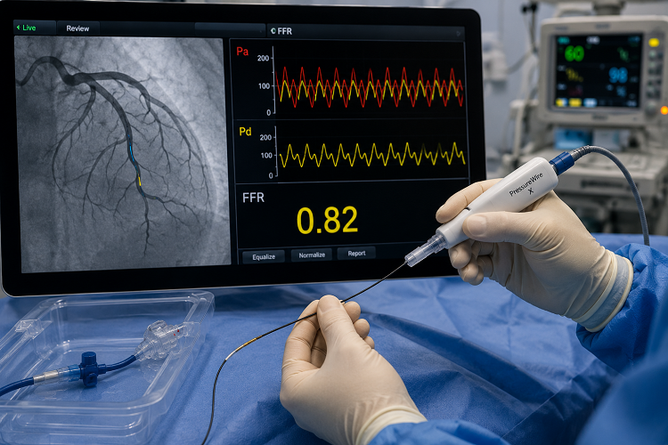
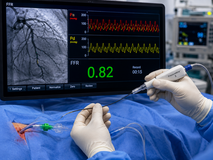
Precision PCI refers to a carefully planned approach to coronary angioplasty where advanced imaging and physiological measurements may be used to understand the artery more accurately before, during and after an intervention.
Traditional angiography provides an outline of the coronary arteries. IVUS and OCT can provide additional information from inside the artery, while FFR can help determine whether a narrowing is significantly affecting blood flow.
By combining anatomical and functional information when clinically appropriate, the cardiologist can make a more informed decision regarding treatment.
IVUS, OCT and FFR provide different types of information. Depending on the patient's condition and coronary anatomy, one or more of these technologies may be used to support treatment planning.
IVUS uses ultrasound from inside the coronary artery to provide detailed information about the vessel, plaque and coronary lesion.
OCT provides high-resolution images from inside the coronary artery and can help assess plaque and the result of stent implantation.
FFR measures pressure differences across a coronary narrowing to help determine whether the blockage is significantly affecting blood flow.
Intravascular Ultrasound (IVUS) uses a small ultrasound transducer positioned inside the coronary artery to provide cross-sectional information about the vessel.
It can help the interventional cardiologist understand vessel dimensions, plaque burden and lesion characteristics, particularly when angiographic images alone may not provide sufficient information.
Optical Coherence Tomography (OCT) uses light-based imaging to produce high-resolution views from inside the coronary artery.
OCT may be particularly useful when detailed visualisation of the coronary lesion or stent result is required.
Fractional Flow Reserve (FFR) is a physiological measurement performed inside the coronary artery to determine whether a narrowing is significantly affecting blood flow to the heart muscle.
FFR can be especially useful when the significance of a coronary narrowing is uncertain and additional information is required before deciding whether intervention is needed.
Advanced imaging and physiological assessment may be considered in selected patients when additional information can help guide coronary intervention.
When angiography alone does not clearly establish the significance of a coronary narrowing.
Useful in selected complex coronary lesions where detailed anatomical assessment is important.
Imaging can help assess stent expansion, apposition and the final PCI result.
Provides additional information to support an individualised coronary treatment strategy.
Modern coronary intervention is increasingly focused on accurate diagnosis, appropriate treatment selection and optimisation of procedural results. IVUS, OCT and FFR can provide complementary information in selected patients.
Better anatomical and physiological understanding for selected coronary interventions.
Dr. Shripad V. Khairnar provides advanced interventional cardiology care with modern coronary assessment techniques. Depending on the patient's condition, IVUS, OCT and FFR may be used to obtain additional anatomical and physiological information and support an individualised treatment strategy.